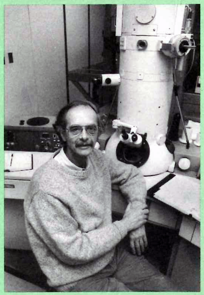
Our longtime colleague, Lee D. Peachey, Ph.D., emeritus Faculty, died in May 2024 at the age of 92, after life rich in experience and academic accomplishment. Peachey grew up in Rochester, NY and attended Lehigh University in Pennsylvania for a B.S. degree in Engineering Physics in 1953. From 1953-1956, he attended the University of Rochester, Department of Radiation Biology and Biophysics, in the Section of Electron Microscopy, working with Dr. Michael L. Watson. Watson was a pioneer of the Electron Microscope, a new instrument at the time allowing unprecedented views into the cell. In 1956, Peachey transferred to The Rockefeller Institute (now Rockefeller University), as the first graduate student of Dr. Keith R. Porter, considered by many the father of cell biology. During this period, Peachey also spent 10 months at Cambridge University with Professor Sir Andrew F. Huxley, a neuroscientist who was awarded the Nobel prize in 1963 for his work on the neural action potential with Hodgkin. At the time, Sir Huxley was focused on the physiology and biophysics of muscle. Peachey received his PhD in June 1959 from Rockefeller, and after a brief period as Research Associate, took up a position as Assistant, then Associate Professor of Zoology at Columbia University, 1960-1965. In 1965, he was recruited to The University of Pennsylvania as Associate Professor of Biochemistry and Biophysics. As fortune would have it, in 1970, he accepted appointment as Chairman of the Department of Biology, serving until 1972, then remaining in the Biology Department as Professor until his retirement in 2000. Despite emeritus status, he worked in his laboratory for many years following retirement, providing experimental and educational advice and guidance, as well as collegiality, to his fellow faculty.
Peachey’s research focus fell under the umbrella of the morphology of cells and tissues for an understanding of how structure relates to physiology and biochemistry of function. Over his career he used a number of different preparations, including muscle for contraction-excitation coupling and mammalian kidney, thyroid hormone effects on mitochondrial morphology, among others. We know him best as a specialist in structure of cells, which initiated with specimen preparation for electron microscopy, quantitative analysis of the data, and evolved to confocal and other types of cutting-edge microscopy as the latter techniques gained in instrumentation and technical breakthroughs. His most cited articles include those characterizing cellular structure in great detail through electron microscopy and sample preparations for the preservation of exceptional tissue detail. A well known and popular colleague on campus, he was active in the Pennsylvania Muscle Institute and collaborated with many colleagues across and outside of the University, being well-respected and well revered in the field for his expertise in muscle biophysics and structural biology. In this capacity, he collaborated and provided expertise as adjunct Professor of the Molecular, Cellular and Developmental Biology Department of the University of Colorado, Boulder from 1969-1984, and International Visiting Professor of Gunma University Medical School, Maebashi, Japan from 1992-1995.
During his academic career, Peachey also engaged in a number of sabbaticals in Cambridge, England, as a Guggenheim Fellow, Fulbright-Hays Fellow, and Overseas Fellow of Churchill College (1967-1968), Fogarty Senior International Fellow in London (1979-1980), and Guest Research Fellow of the Royal Society of London, University College London (1986-1987). He volunteered his time in positions in the Biophysical Society (President from 1981-1982), the Electron Microscopy Society of America (President 1982), the International Society for Pure and Applied Biophysics (President 1987-1990), among others, and was actively engaged in grant service for the National Institutes of Health, journal reviewing, and served the City of Philadelphia on the Mayor’s Science and Technical Advisory Council. He also spent time at Woods Hole, and hosted Dr. Keith Porter in his laboratory as emeritus from 1988 until Porter’s death in 1997. He also served as Trustee for the Keith R. Porter Endowment for Cell Biology from 1981-2004.
Many of us in the Department, however, know him best for his devotion to securing high quality cutting edge instrumentation for microscopy needs in the Department and the University, and as a fervent devotee of the education mission of the University. He taught a number of courses early on, including Biology 103: Molecular and Cellular Biology and Biology 6: Human Biology, and engaged in voluntary technical courses in microscopy technique to members of the Department and University throughout his career. But to those of us in neuroscience, his legacy course is neurobiology, now known as Biol 2110: Molecular and Cellular Neurobiology. He was deeply devoted to helping newly hired Assistant Professors master the lectures and materials, active in collaborating with them on the structure, set up and direction of the course, and was a keen supporter of the establishment and instrumentation of a laboratory component to this popular fundamental campus neuroscience course. Through these activities, we came to know him as a devoted colleague, intellectually engaged associate, and thoughtful faculty partner with a critical and helpful eye, and with a warm sense of humor.
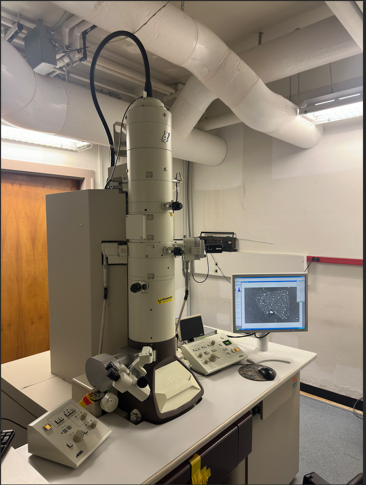
Below: A Joel electron microscope of the type Lee Peachey, Ph.D., mastered. An enormous machine that allows visualization of the microscopic ultrastructural elements of cells and tissues. This machine is housed in the basement of Leidy Laboratory in the same room where Lee Peachey’s machine was housed, but is a more recent model. Image courtesy of Changsong Yang and Prof. Tatyana Svitkina, by Prof. Nancy Bonini, Department of Biology.